If you′re living with GA, or have AMD and notice any vision changes, it′s important to talk to an eyecare professional, such as your optometrist or an ophthalmologist (specialist eye doctor).
The Impact of Geographic Atrophy
 GA affects approximately 75,000 people in Australia and 5 million globally.

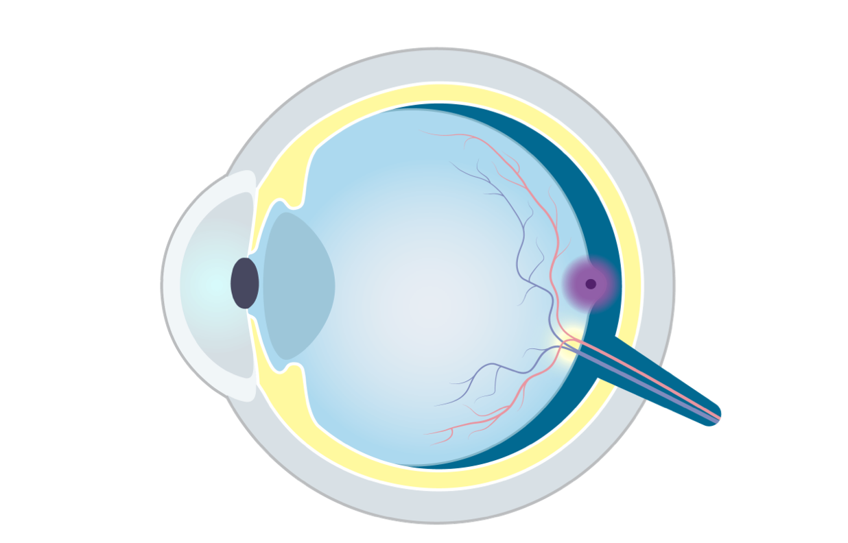
Understanding the eye
Your eyes help you navigate the world and connect with the people and things you love.
Vision happens when your eyes turn light into signals your brain can recognise.
Learning about how the eye works will help you understand how eye diseases like geographic atrophy (GA) can affect the eye.

View transcriptClose transcript
If you are affected by geographic atrophy, as a patient, a family member or a caregiver, it is important to understand how we see.
For someone with healthy eyes, seeing comes naturally. But how does the eye actually work? Let's take a look at how we see.
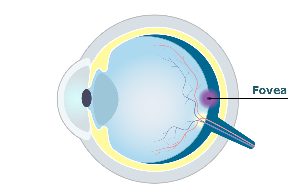
Basically, the eye looks like a sphere.
In the front part of this sphere, we′ve got a typically brown, blue or green coloured circle - the iris. It is coloured due to its pigments. It regulates the amount of light that enters the eye through the pupil by controlling its size.
The pupil is the black opening in the centre of the iris. It allows light to enter the inside of the eye reaching the retina.
This is the vitreous humour. It′s a clear gel that fills the space between the lens and the retina at the back of the eye. Along with maintaining the shape of the eye, the vitreous humour helps absorb shocks to the eye and keeps the retina properly connected to the back wall of the eye.
Here is the lens, a transparent disc behind the iris and the pupil. The lens helps to focus the light and project it onto the retina. The lens enables the eye to see sharply both near and far.
This part right in the front of the eye is called the cornea. It′s the clear dome covering the front of the eye including the iris and the pupil. The cornea helps the eye focus the light, so objects look sharp and clear.
Then there is the sclera. The sclera is the white outer layer of the eye, contrasting with the coloured iris. The sclera provides protection and form to the eye.
Let′s look at the back of the eye: There we find the retina, a light-sensitive tissue lining the inner back of the eye.
And there′s the optic nerve. This bundle of 100 million nerve fibers carries visual messages as electric impulses from the retina to the visual cortex of the brain.
This small area of the retina is called the macula. It has a diameter of around 5.5 mm. The macula is responsible for the central, high-resolution and colour vision.
In the middle we find a small pit we call the fovea responsible for sharp central vision used for reading and driving.
Now, how does this all work together, so we can actually see what we see?
Light enters through the cornea and the pupil into the eye. It then travels through the vitreous humour, the clear gel, and falls onto the retina at the back of the eye.
Here, it′s turned into electric impulses. And these impulses will be carried to the brain by the optic nerve.
In summary, the eye is an organ where different parts work together in complex ways to make us see.
Let′s take a look at how this transmission works in more detail.
To do this we have to take a closer look at the macula. Remember? That′s the part at the back of the eye that is responsible for central vision.
When light beams enter the eye and reach the retina, they activate photoreceptors, which convert the light stimulus into electrical signals.
These electrical signals are sent by the photoreceptors to the bipolar cells, and subsequently activate the retinal ganglion cells.
From here the signals travel through the nerve cords of the retinal ganglion cells to the optic nerve, which transmits the information to the visual cortex in the brain.
In summary, the retina converts light energy into electric signals.
To understand how light is transformed into electric signals, we take a closer look at those photoreceptors in the retina.
There are about 126 million receptors in each eye. Six million in the form of cones, and 120 million shaped as rods. The function of the cones is to detect daylight, colours and fine details, and they mainly manage the central vision. The rods, on the other hand, are good for detecting large moving objects, grey scale, and nightlight. They can process less detail and are there mostly for the peripheral vision.
In summary, rods and cones are photoreceptors in the retina converting light beams into electric impulses.
The highest concentration of photoreceptors in the retina is in the macula. It′s the sensitive area, about 5.5 mm in diameter, needed for the central vision.
Here we see in closer details the pit in its centre that is called the fovea. It provides the sharpest and most detailed vision because of the high concentration of cones. Also, the light can pass directly onto the photoreceptors as the inner retinal layers are pushed to either side.
In summary, the fovea is the spot in the macular where vision is the sharpest.
Let's recap the process of seeing in brief:
Light passes through the iris and the pupil. It is focused by the cornea and the lens onto the retina. In the retina, it is focused in the region of the macula. Photoreceptors convert the light into electric impulses that travel through the optic nerve to the visual cortex in the brain.
That's the way we see.
Click the arrow to explore each part of the eye
Swipe the arrow to explore each part of the eye
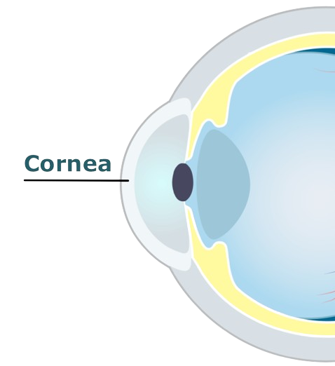
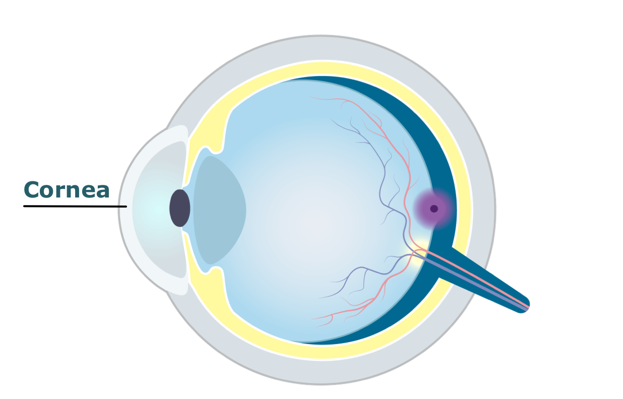
The cornea is clear and dome-shaped. It helps focus light as it enters the front of the eye.
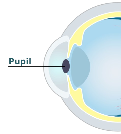
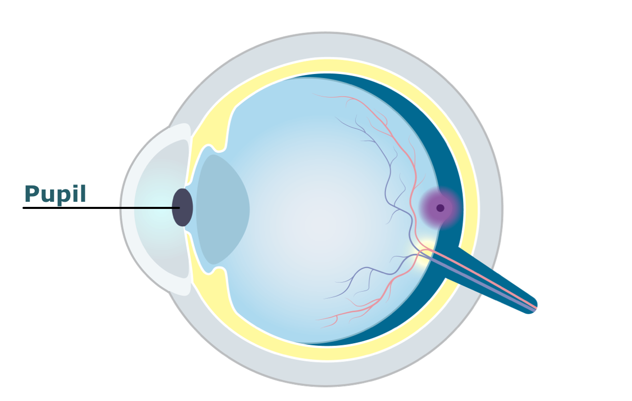
The pupil is the entryway for light. This hole in the centre of your eye helps manage how much light is reaching the eye.
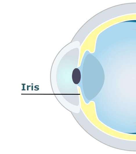
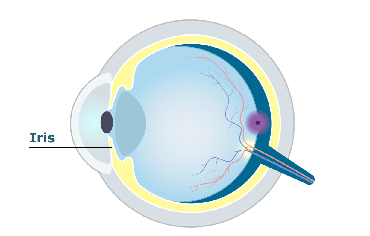
The iris is a colourful ring that surrounds the pupil. Muscles in the iris expand and contract to control the amount of light entering the eye.
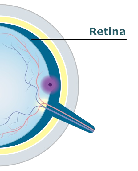
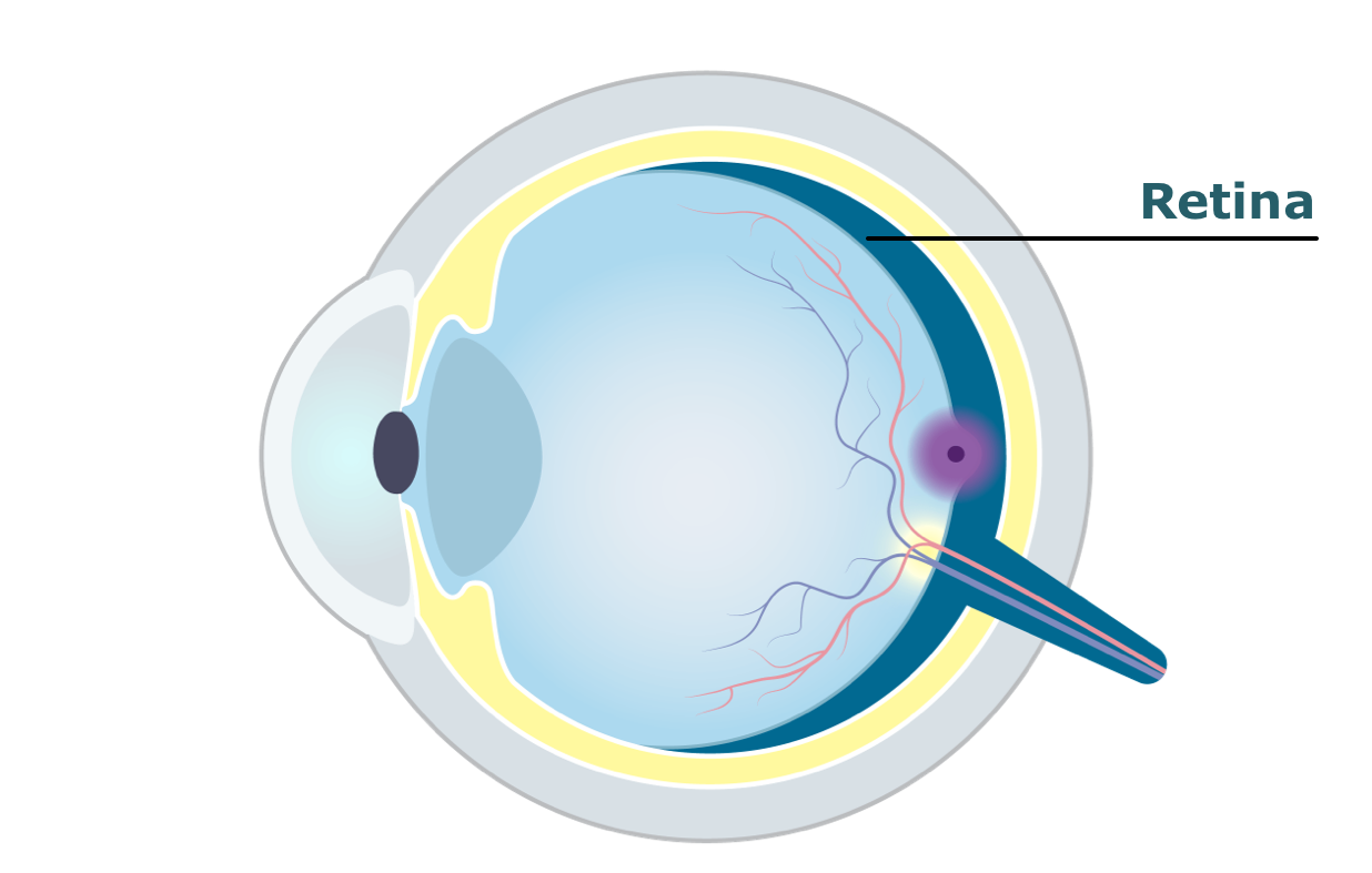
The retina lines the back wall of the eye. It turns light into signals that help the brain recognise what you are seeing.
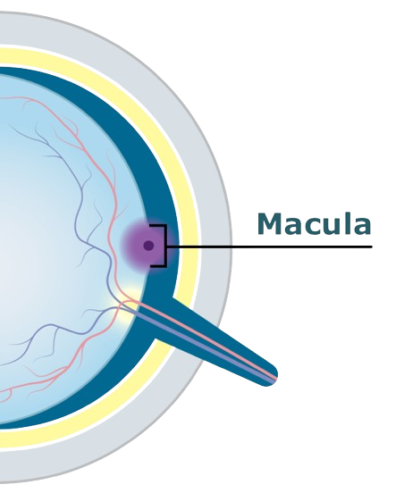

The macula is a very tiny section of the retina. It helps you see in detail.
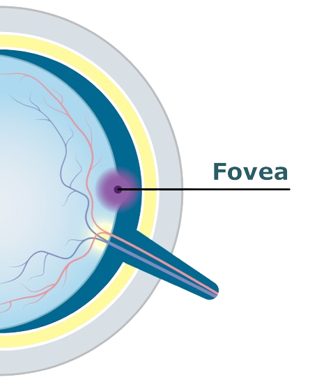
The fovea is the centre of the macula. It helps create sharp, clear images of the things you are focusing on.
How GA impacts vision
GA can affect vision in different ways. You may not notice vision changes when looking at an eye chart. However, everyday activities such as reading and driving at night, may become harder. This happens as patches of damage, called lesions, grow larger.
TapHover over the images to see some signs and symptoms of GA*
Difficulty seeing in the dark


Hazy or blurred vision (less sharp or detailed)
Straight lines appear crooked

A small but growing blurry spot in the centre of vision
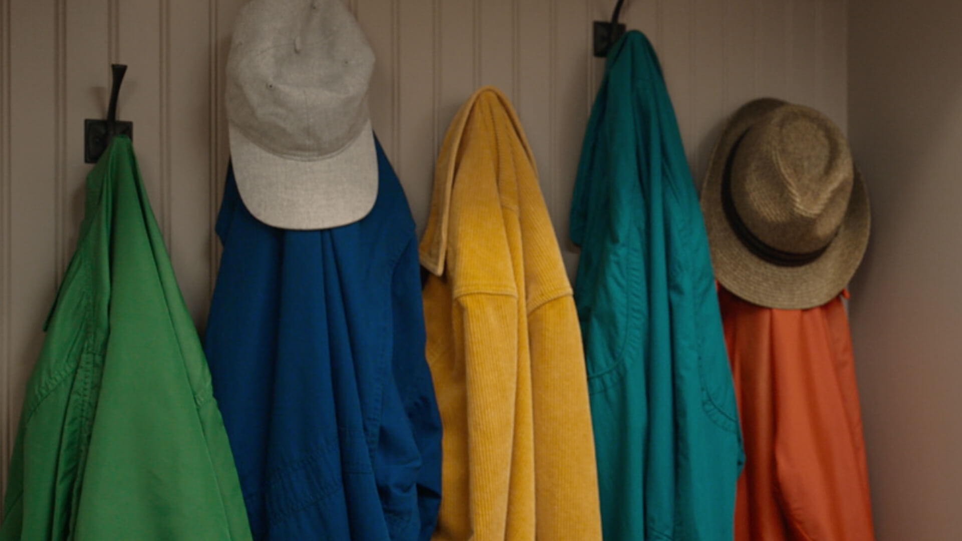
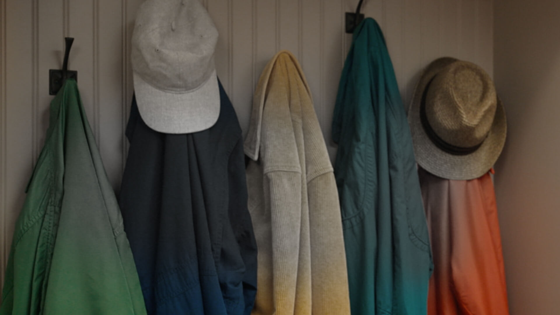
Colours seem dull or washed out
*For illustration purposes only. Vision impairment due to GA may vary.
See how GA changes vision over time
Click to experience the progression of GA over time.†
See how GA changes vision over time
Swipe to experience the progression of GA over time.†

†For illustrative purposes only. Vision impairment due to GA may vary.
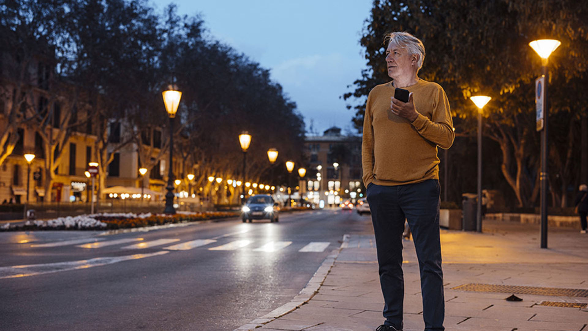
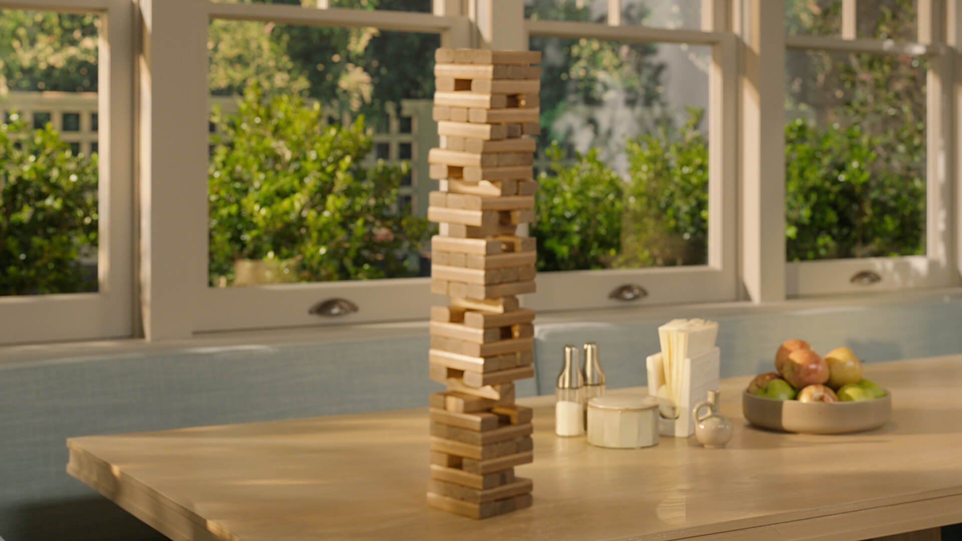
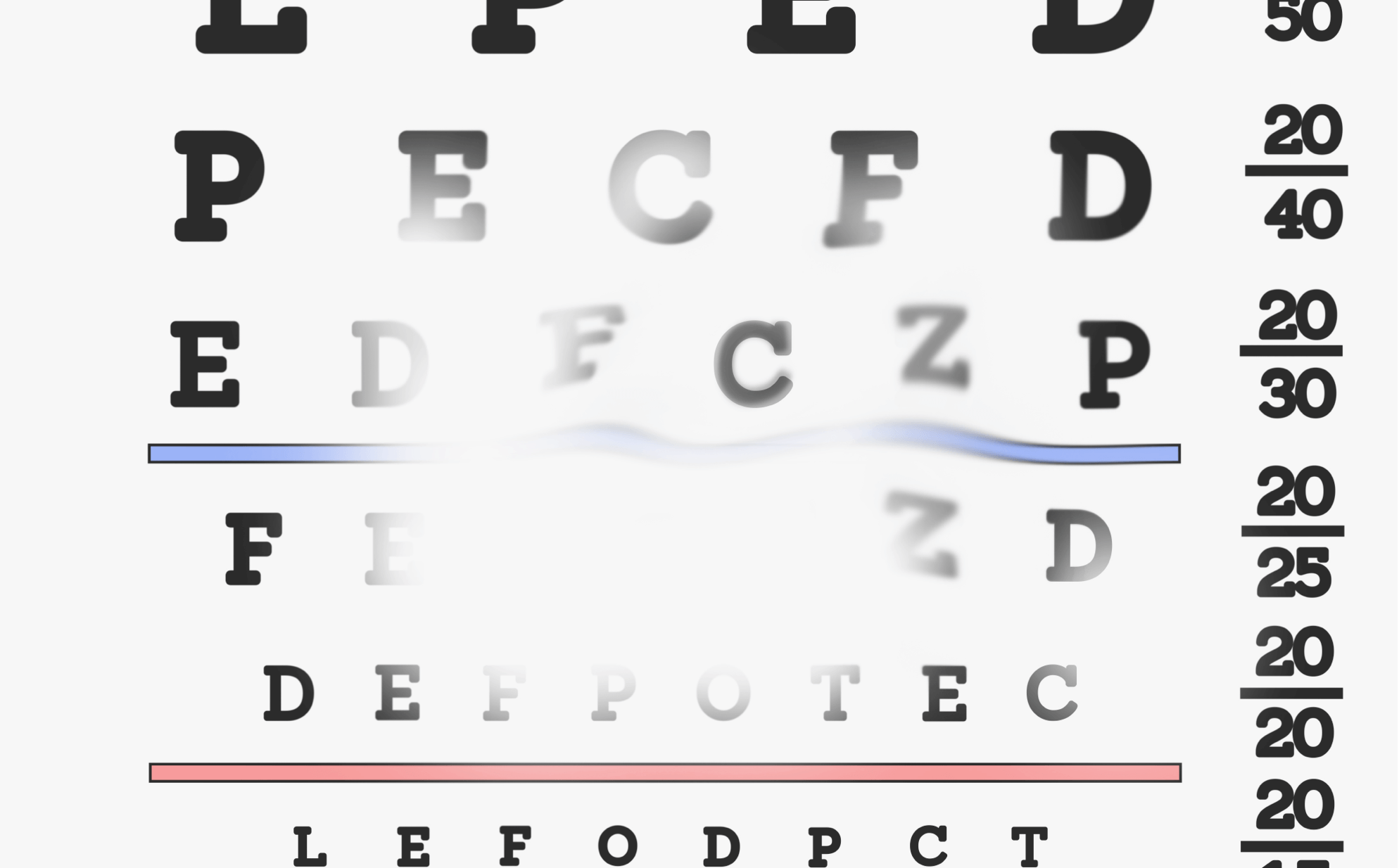
At diagnosis
This image shows the vision of a 70-year-old woman who has just been diagnosed with GA in both eyes. Her eyes have multiple lesions in the area surrounding the fovea.
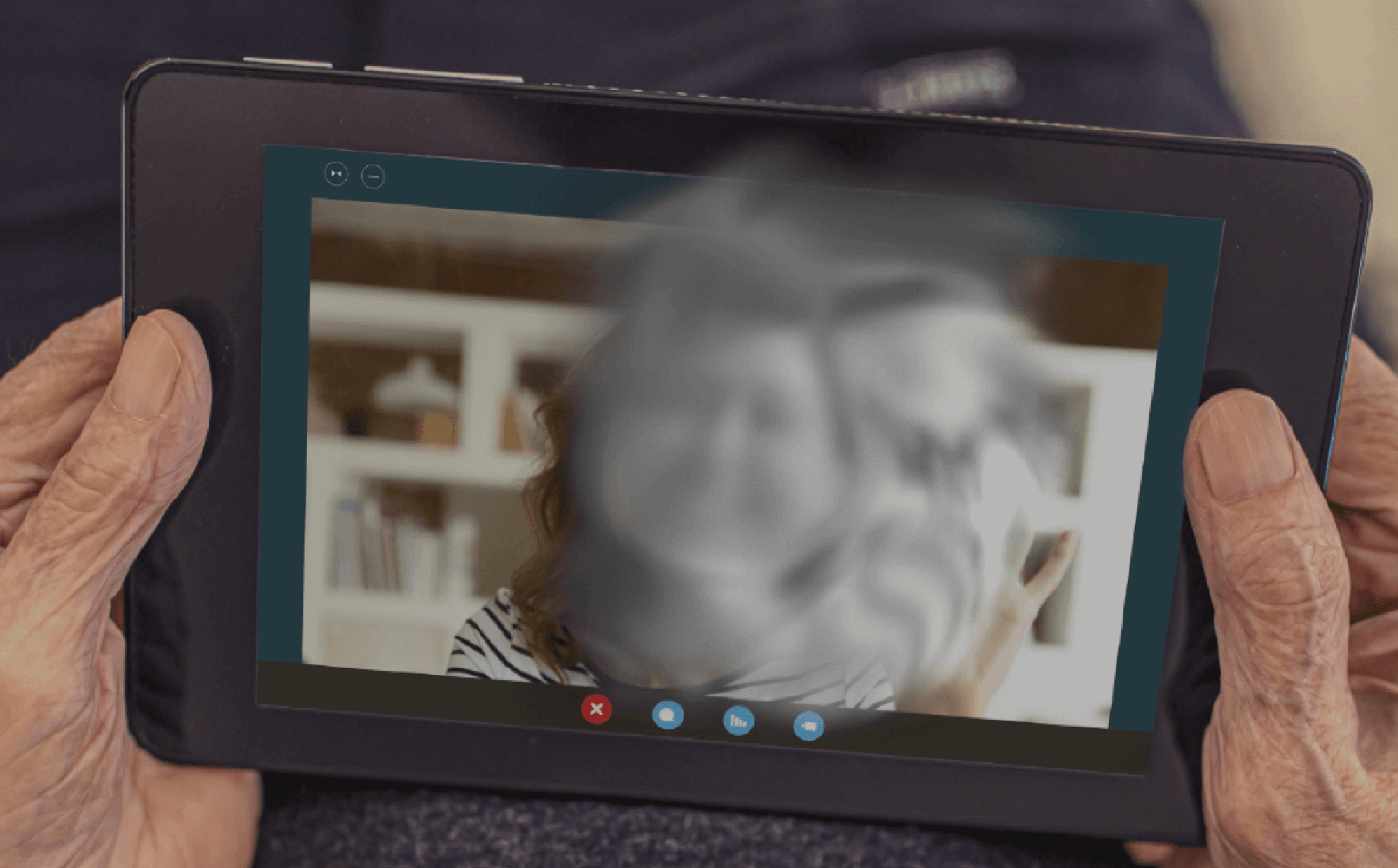
†For illustrative purposes only. Vision impairment due to GA may vary.
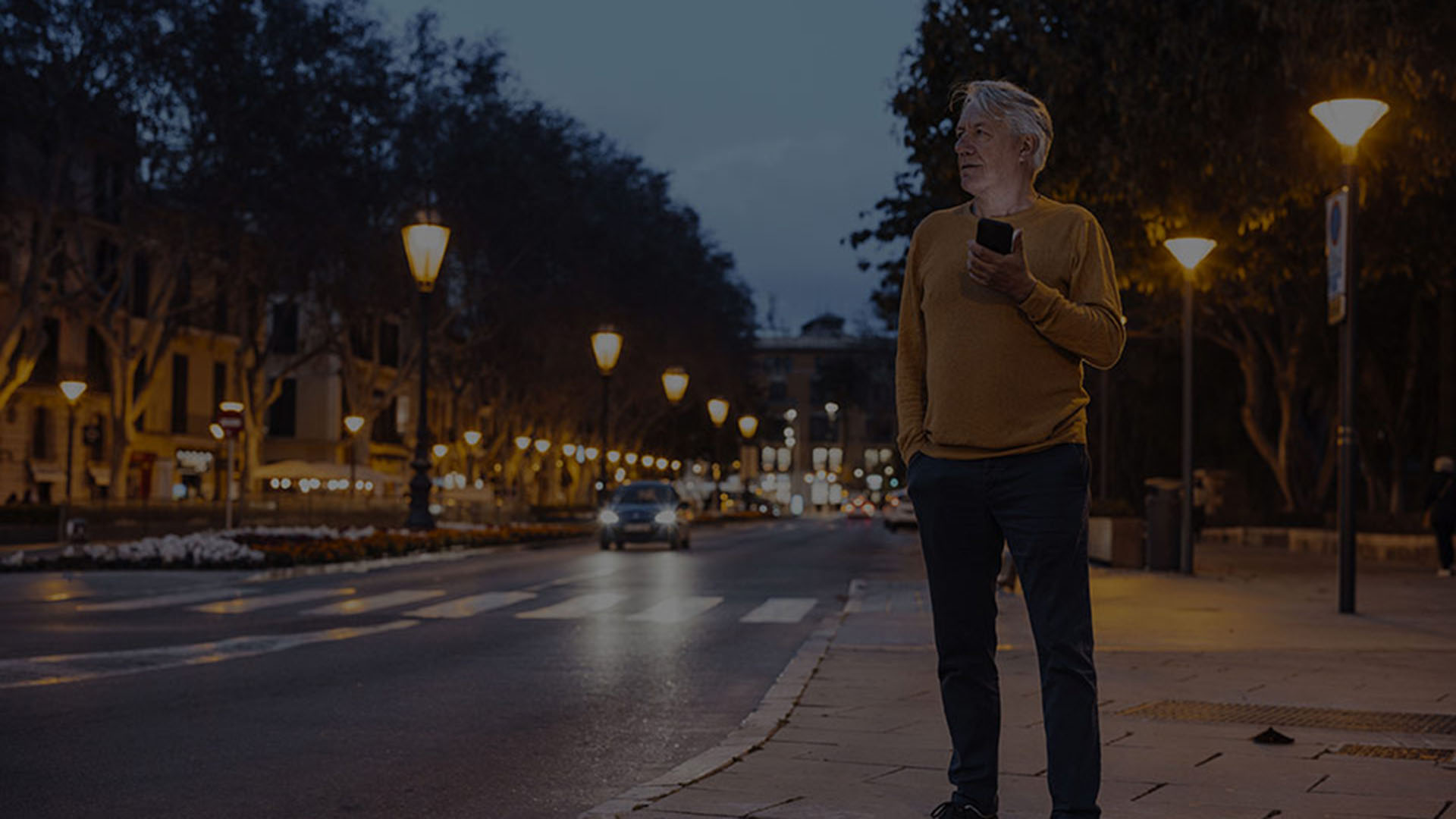
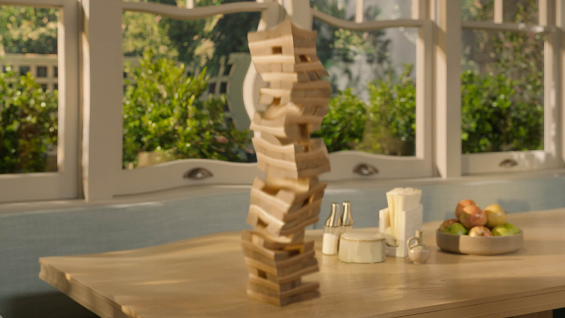
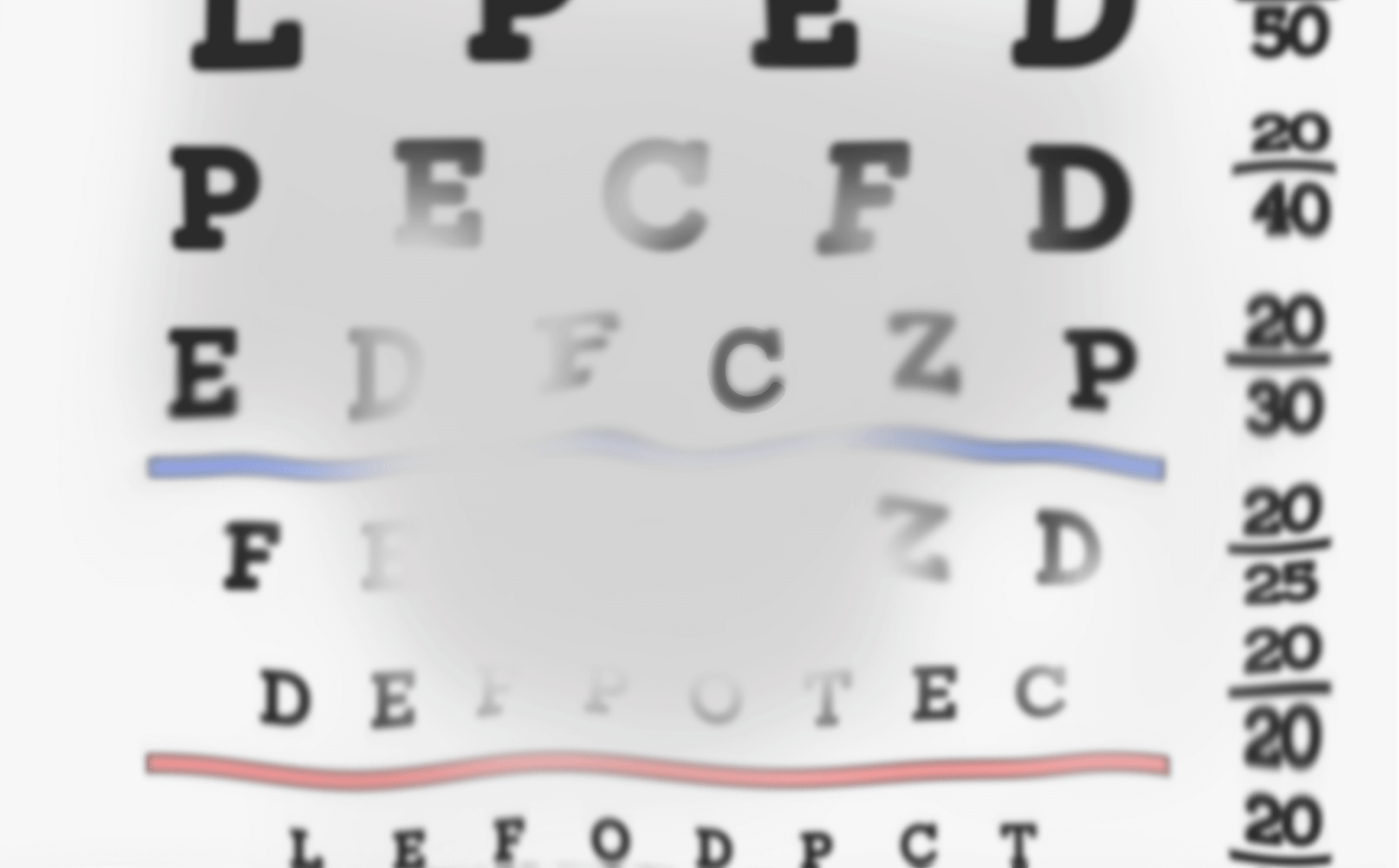
+2 years
GA has progressed in both of the patient′s eyes. The faces of loved ones have become harder to see. Driving and other daily activities may become unsafe as lesions grow larger.
†For illustrative purposes only. Vision impairment due to GA may vary.
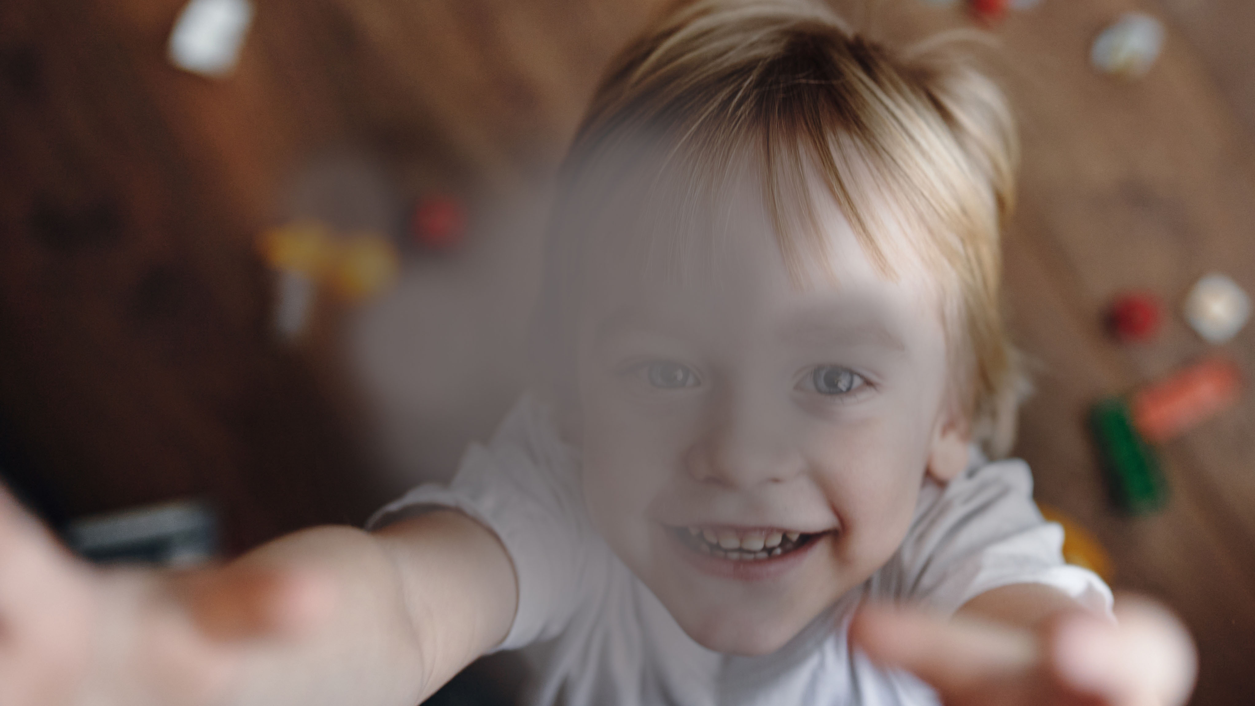
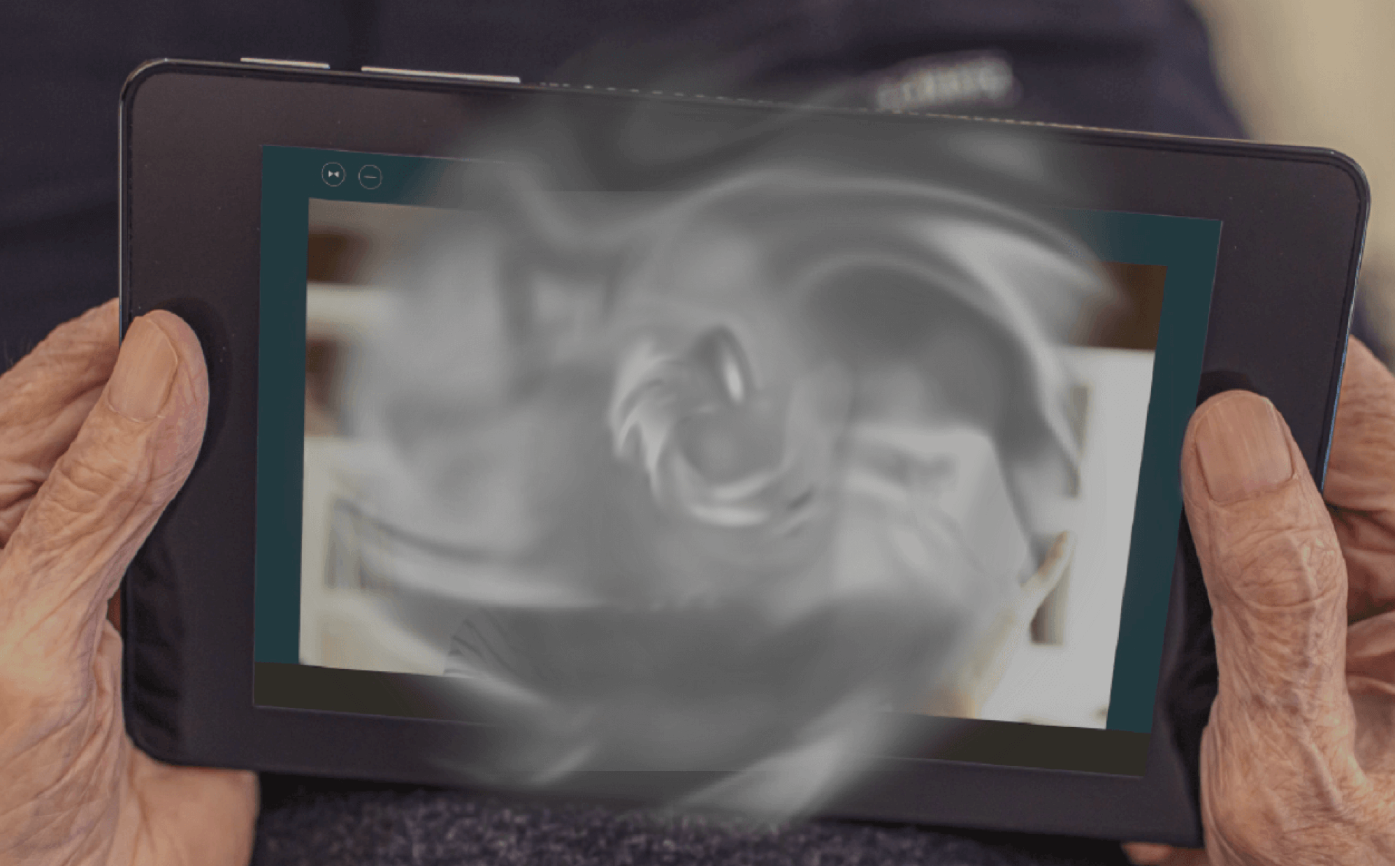
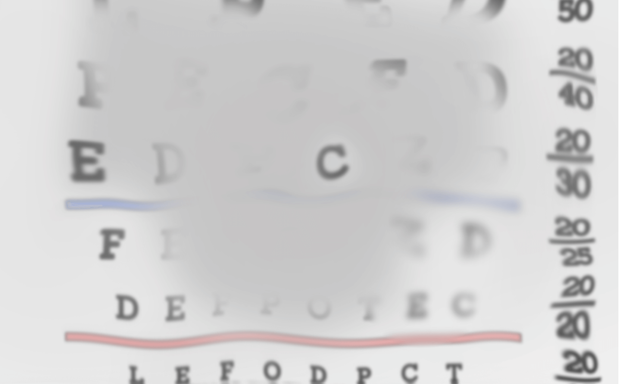
+6 years
Now, with increased progression in both eyes, reading, the ability to live independently, and overall quality of life have all been impacted by vision loss due to GA.
†For illustrative purposes only. Vision impairment due to GA may vary.
At diagnosis
This image shows the vision of a 70-year-old woman who has just been diagnosed with GA in both eyes. Her eyes have multiple lesions in the area surrounding the fovea.

†For illustrative purposes only. Vision impairment due to GA may vary.
+2 years
GA has progressed in both of the patient′s eyes. Reading has become more difficult. Driving and other daily activities may become unsafe as lesions grow larger.

†For illustrative purposes only. Vision impairment due to GA may vary.
+6 years
Now, with increased progression in both eyes, the faces of loved ones, the ability to live independently, and overall quality of life have all been impacted by vision loss due to GA.
†For illustrative purposes only. Vision impairment due to GA may vary.
At diagnosis
This image shows the vision of a 70-year-old woman who has just been diagnosed with GA in both eyes. Her eyes have multiple lesions in the area surrounding the fovea.
†For illustrative purposes only. Vision impairment due to GA may vary.
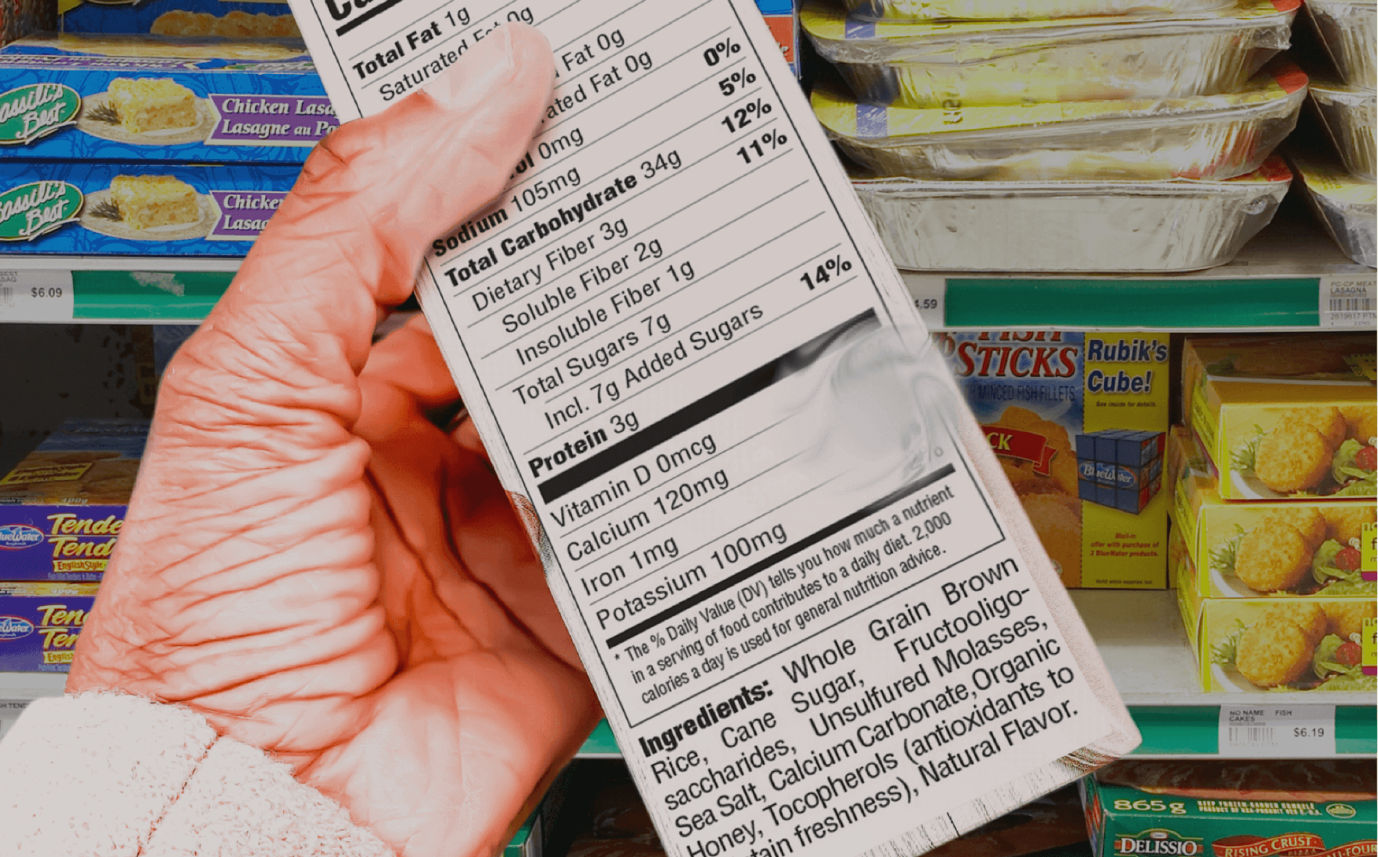
+2 years
GA has progressed in both of the patient′s eyes. Letters have become harder to see. Driving and other daily activities may become unsafe as lesions grow larger.
†For illustrative purposes only. Vision impairment due to GA may vary.
+6 years
Now, with increased progression in both eyes, reading, the ability to live independently, and overall quality of life have all been impacted by vision loss due to GA.

Contributing factors for GA
Below is a list of things that may contribute to GA:
Age

Family history (the genes you inherit play ~70% of the role in GA development)

Diet high in fatty foods
High blood pressure

History of smoking
Low physical activity

Obesity
UV exposure
Be prepared to talk to your eye doctor or optometrist about changes in vision and potential signs of GA.
Are you an eyecare professional?
You can now access a dedicated resource on geographic atrophy